预约演示
更新于:2026-08-22
AVN-944
更新于:2026-08-22
概要
基本信息
在研机构- |
最高研发阶段终止临床2期 |
首次获批日期- |
最高研发阶段(中国)- |
特殊审评- |


登录后查看时间轴
结构/序列
分子式C25H27N5O5 |
InChIKeyGYCPCOJTCINIFZ-JXFKEZNVSA-N |
CAS号915951-68-7 |
Sequence Code 15387629

关联
3
项与 AVN-944 相关的临床试验NCT00923728
A Phase 1 Trial of AVN944, an IMPDH Inhibitor, in Adults With Advanced Stage Solid Tumors
NCT00493441
A Phase II Study of AVN944 in Combination With Gemcitabine for the Treatment of Pancreatic Cancer
NCT00273936
A Phase I Trial of AVN-944 in Patients With Advanced Hematologic Malignancies
100 项与 AVN-944 相关的临床结果
登录后查看更多信息
100 项与 AVN-944 相关的转化医学
登录后查看更多信息
100 项与 AVN-944 相关的专利(医药)
登录后查看更多信息
15
项与 AVN-944 相关的文献(医药)2026-03-04ANTIMICROBIAL AGENTS AND CHEMOTHERAPY
Exploring
T. cruzi
IMPDH as a promising target through Chagas Box screening and AVN-944 inhibition
Article
作者: Lobo-Rojas, Angel ; Faria, Jessica D. N. ; Eufrasio, Amanda G. ; Souza, Gabriel T. D. ; Marchese, Letícia ; Cordeiro, Artur T. ; Catelli, Michelle F.
ABSTRACT:
Chagas disease, caused by
Trypanosoma cruzi
, remains a leading cause of heart failure in Latin America, with current treatments limited to acute-phase efficacy, significant toxicity, and emerging resistance. Inosine monophosphate dehydrogenase (IMPDH) is an essential enzyme in guanine nucleotide salvage pathway and represents a promising alternative target. Here, we combined computational screening, biochemical and cell-based phenotypic assays that support
T. cruzi
IMPDH (
Tc
IMPDH) as a druggable target and identify repurposing opportunities among clinical-stage inhibitors. Using Tanimoto similarity scoring against the library of 222 Chagas Box compounds, we identified TCMDC-143376 as uniquely similar to the clinical IMPDH inhibitors merimepodib and AVN-944. Phylogenetic analysis and multiple sequence alignment confirmed conservation of both catalytic and allosteric residues—drawn from
T. foetus
and
T. brucei
structures—within
Tc
IMPDH. Recombinant
Tc
IMPDH kinetics revealed Michaelis constants of 155 µM for IMP and 292 µM for NAD
+
. Biochemical IC
50
assays showed submicromolar inhibition by AVN-944 (0.20 µM), (S)-Merimepodib (0.21 µM), and (R)-Merimepodib (0.37 µM). In H9c2 cardiomyoblasts infected with intracellular amastigotes, AVN-944 achieved the lowest EC
50
(0.4 µM), outperforming benznidazole (EC
50
= 3.0 µM) and other inhibitors. Our findings support
Tc
IMPDH as a promising alternative drug target for Chagas disease and position AVN-944 as a compelling candidate to evaluate this therapeutic strategy in animal models.
2025-08-01MOLECULAR DIVERSITY
Cangrelor and AVN-944 as repurposable candidate drugs for hMPV: analysis entailed by AI-driven in silico approach
Article
作者: Henna, Fathimath ; John, Levin ; Raju, Rajesh ; Jayanandan, Abhithaj ; Thomas, Sonet Daniel ; Thaikkad, Amritha
Human metapneumovirus (hMPV) primarily causes respiratory tract infections in young children and older adults. According to the 2024 Human Pneumonia Etiology Research for Child Health (PERCH) study, hMPV is the second leading common cause of pneumonia in children under five in Asia and Africa. The virus encodes nine proteins, including the essential Fusion (F) and G glycoproteins, which facilitate entry to the host cells. Currently, there are no approved vaccines or antiviral treatments for hMPV; supportive care is the primary way it is managed. Hence, this study focuses on the F protein as a therapeutic target to find a repurposable drug to fight hMPV. Refolding of the F protein and its binding to heparan sulfate enable hMPV infection. Heparin sulfate is important for hMPV binding, and we have found that cangrelor and AVN 944 can prevent the fusion of membranes. We developed a deep learning-based pharmacophore to identify potential drugs targeting hMPV, from which we could narrowed a list of 2400 FDA-approved drugs and 255 antiviral drugs to 792 and 72 drugs, respectively. We then conducted quantitative validation using the ROC curve. Further virtual screening of the drugs was performed, leading us to select the one with the highest docking score. The validation of the deep learning prediction in virtual screening Pearson correlation was done. Further, the MD simulation of these drugs confirmed that the protein-drug complex stability remained in dynamic condition. Further, the stability of protein-drug complexes than unbound protein was confirmed by Free Energy Landscape and Dynamic Cross Correlation Matrices. Further in vitro and in vivo experiments need to determine the efficacy of the identified candidates.
2025-04-01ACTA PHARMACOLOGICA SINICA
IMPDH inhibitors upregulate PD-L1 in cancer cells without impairing immune checkpoint inhibitor efficacy
Article
作者: Yang, Bo ; Wu, Hong-Hai ; Li, Jia-Yi ; Zhang, Jie ; He, Qiao-Jun ; Wang, Long-Sheng ; Zheng, Ming-Ming ; Ding, Ling ; Jiang, Ke-Fan ; Guo, Hong-Jie
Tumor cells are characterized by rapid proliferation. In order to provide purines for DNA and RNA synthesis, inosine 5'-monophosphate dehydrogenase (IMPDH), a key enzyme in the de novo guanosine biosynthesis, is highly expressed in tumor cells. In this study we investigated whether IMPDH was involved in cancer immunoregulation. We revealed that the IMPDH inhibitors AVN944, MPA or ribavirin concentration-dependently upregulated PD-L1 expression in non-small cell lung cancer cell line NCI-H292. This effect was reproduced in other non-small cell lung cancer cell lines H460, H1299 and HCC827, colon cancer cell lines HT29, RKO and HCT116, as well as kidney cancer cell line Huh7. In NCI-H292 cells, we clarified that IMPDH inhibitors increased CD274 mRNA levels by enhancing CD274 mRNA stability. IMPDH inhibitors improved the affinity of the ARE-binding protein HuR for CD274 mRNA, thereby stabilizing CD274 mRNA. Guanosine supplementation abolished the IMPDH inhibitor-induced increase in PD-L1 expression. In CT26 and EMT6 tumor models used for ICIs based studies, we showed that despite its immunosuppressive properties, the IMPDH inhibitor mycophenolate mofetil did not reduce the clinical response of checkpoint inhibitors, representing an important clinical observation given that this class of drugs is approved for use in multiple diseases. We conclude that PD-L1 induction contributes to the immunosuppressive effect of IMPDH inhibitors. Furthermore, the IMPDH inhibitor mycophenolate mofetil does not antagonize immune checkpoint blockade.
1
项与 AVN-944 相关的新闻(医药)100 项与 AVN-944 相关的药物交易
登录后查看更多信息
外链
| KEGG | Wiki | ATC | Drug Bank |
|---|---|---|---|
| - | - | - |
研发状态
10 条进展最快的记录, 后查看更多信息
登录
| 适应症 | 最高研发状态 | 国家/地区 | 公司 | 日期 |
|---|---|---|---|---|
| 胰腺腺癌 | 临床2期 | 美国 | 2007-06-01 | |
| 自身免疫性疾病 | 临床2期 | - | - | |
| 急性白血病 | 临床1期 | 美国 | 2006-01-01 | |
| 慢性白血病 | 临床1期 | 美国 | 2006-01-01 | |
| 霍奇金淋巴瘤 | 临床1期 | 美国 | 2006-01-01 | |
| 多发性骨髓瘤 | 临床1期 | 美国 | 2006-01-01 | |
| 巨球蛋白血症 | 临床1期 | 美国 | 2006-01-01 | |
| 血液肿瘤 | 临床1期 | 美国 | - | |
| 晚期恶性实体瘤 | 临床阶段不明 | 美国 | 2009-04-01 |
登录后查看更多信息
临床结果
临床结果
适应症
分期
评价
查看全部结果
登录后查看更多信息
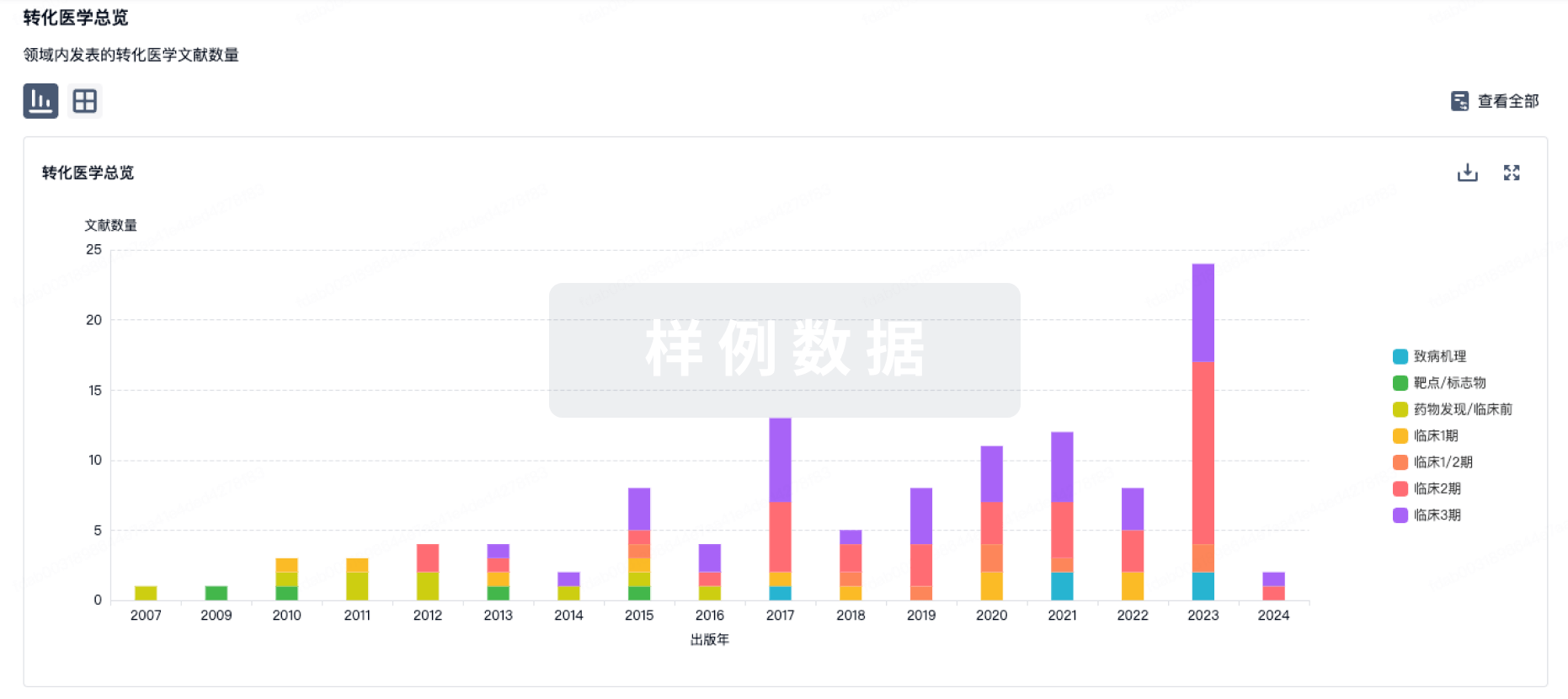
转化医学
使用我们的转化医学数据加速您的研究。
登录
或
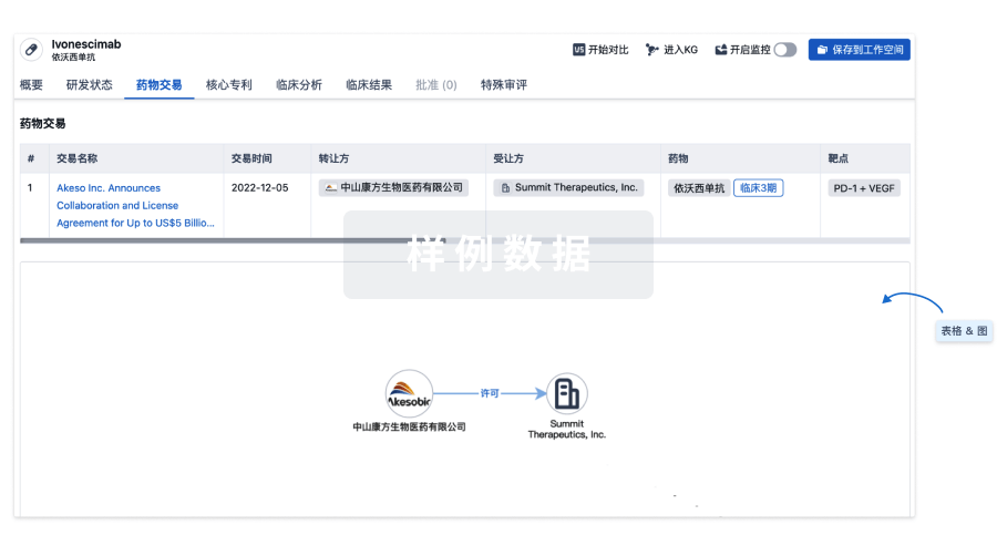
药物交易
使用我们的药物交易数据加速您的研究。
登录
或
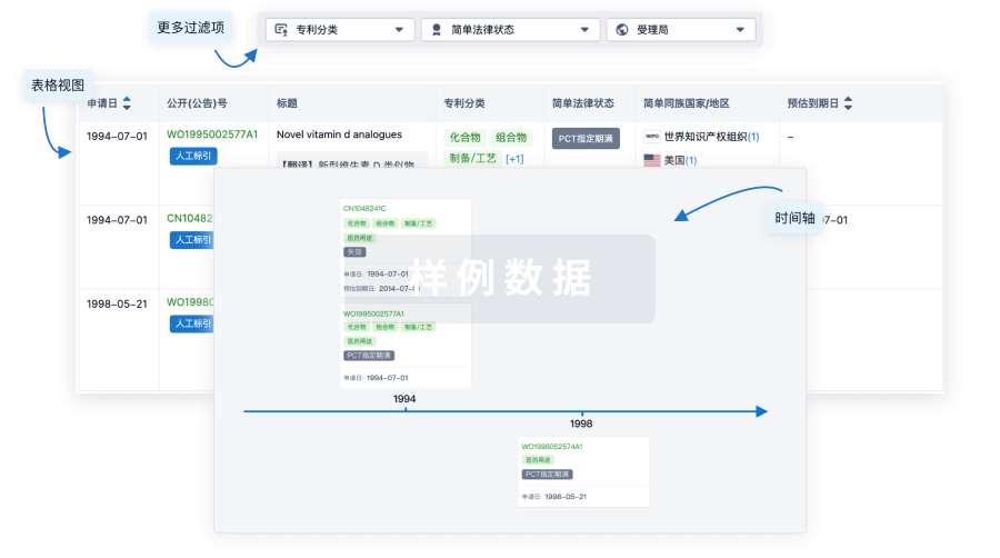
核心专利
使用我们的核心专利数据促进您的研究。
登录
或
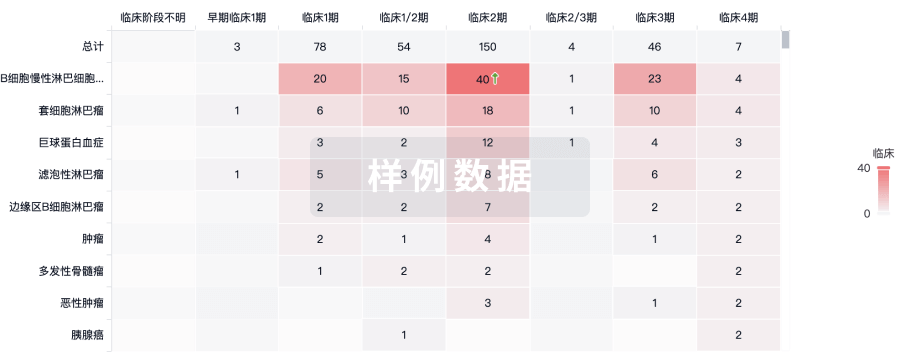
临床分析
紧跟全球注册中心的最新临床试验。
登录
或
批准
利用最新的监管批准信息加速您的研究。
登录
或
特殊审评
只需点击几下即可了解关键药物信息。
登录
或
提出任何生物医药研究问题
让每一步生物医药决策都精准制胜
基于十亿级生物数据与全球情报的精准 AI,赋能早期药物发现
立即开始免费试用!
智慧芽新药情报库是智慧芽专为生命科学人士构建的基于AI的创新药情报平台,助您全方位提升您的研发与决策效率。
立即接入生物医药数据包
智慧芽新药库数据也通过智慧芽开放平台,以MCP或API服务形式对外开放。立即使用我们的MCP服务定制您的LLM智能体。
生物序列数据库
生物药研发创新
免费使用
化学结构数据库
小分子化药研发创新
免费使用